Top of Site/Media
Hemophagocytic Mastocytoma in Cat

Jpn J Vet Dermatol 2009, 15 (2): 75-78
Hemophagocytic Mastocytoma with
Multiple Cutaneous Tumors in a Cat
Shiro Tachikawa: Tachikawa Animal Hospital
Abstract: A 12-year-old neutered male domestic cat was presented with vomiting, lethargy. The cat had 5-month history of multiple cutaneous tumors which had developed as a single cutaneous mass on a hind limb diagnosed as benign mastocytoma by a home veterinarian. Blood smear examination revealed peripheral mastocytosis. Hepatosplenomegaly was not evident by ultrasonographic examination, however cytological assessment of aspirates from the spleen and liver revealed many neoplastic mast cell showing frequent erythrophagocytosis. From these findings the cat was diagnosed as visceral mastocytoma, and then treated with fammotidine and prednisolone. Clinical symptoms had tentatively improved, but anemia had been exacerbated. Thereafter, the neoplastic mast cells in the peripheral blood frequently showed engulfment of erythrocytes and platelets. The cat died on the 26th day.
Key words: hemophagocytic mastocytoma, visceral mastocytoma, cat
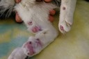
Fig. 1. Multiple cutaneous mast cell tumors in the hind limbs.
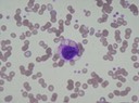
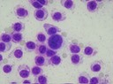
Fig. 2. Cytological features of the fine-needle aspirate of the cutaneous mass. Neoplastic mast cells showing anisocytosis and anisokaryosis contain moderate amount of fine granules.
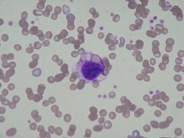
Fig. 3. Neoplastic mast cell showing erythrophagia in the peripheral blood.
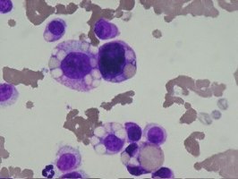
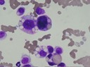
Fig. 4. Cytological features of fine-needle aspirate of the spleen. Note erythrophagocytosis by neoplastic mast cells with anisocytosis, anisokaryosis and a few fine granules.
http://www.jstage.jst.go.jp/article/jjvd/15/2/15_75/_article